top of page


Lymph Node Biopsy

Lymph Node Biopsy
Lymph node biopsy
It is not uncommon for patients to develop enlargement of the lymph nodes in the chest. When lymph nodes become enlarged it is termed lymphadenopathy.

The lymph nodes collect lymph, which is type of fluid that circulates in the body.
The lymph system is almost like a form of cleansing system.
There are lymph nodes in many areas of the body, typically in central locations - that is, you don't have lymph nodes in your fingers, but the lymphatic system from the fingers and arm drains into lymph nodes in the armpit.
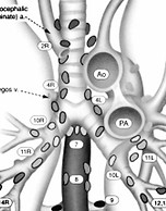
The lymph nodes in the chest drain the lungs and heart and esophagus. The middle of the chest is called the mediastinum, so the lymph nodes in the middle of the chest are called mediastinal lymph nodes. A patient with enlarged mediastinal lymph nodes is said to have mediastinal lymphadenopathy.
Mediastinal lymphadenopathy can occur for several reasons. Causes include:
- Infection, such as bacterial, viral, TB, fungal
- Lymphoma
- Cancer
- Autoimmune process, such as sarcoidosis
- Inflammation
If a patient has no symptoms and exhibits mediastinal lymphadenopathy on a scan, it may make sense to follow the appearance of the lymph nodes on additional scans.
If the nodes are very large or getting larger or if the patient has symptoms - such as shortness of breath or pain or fevers - then a biopsy may be needed to help determine the cause.
A biopsy of the mediastinal lymph nodes can be performed several different ways, including:


CME
EBUS

ROBOTIC

EBUS stands for EndoBronchial Ultrasound.
EBUS is a procedure, not a surgery. It is usually performed by a pulmonologist (lung doctor). It involves guiding a small ultrasound probe down the trachea.
The probe can help the pulmonologist find lymph nodes. The probe allows for a tiny needle to sample the lymph nodes.
EBUS is one of the least-invasive ways to get lymph node tissue for biopsy.
Patients can usually go home the same day as EBUS.
Because the needles are tiny, sometimes a biosy is not successful with EBUS.
Certain locations in the chest are easier to get to with EBUS than others. There may be some lymph nodes that cannot be reached with EBUS.
Dr. Pool does not perform EBUS. He works with pulmonologists who are highly skilled at EBUS, including
Dr. Tony Ortegon.

Dr. Ortegon

CME stands for Cervical Mediastinal Exploration.
CME involves a small incision in the lower neck, which allows access behind the breastbone into the mediastinum.
Small pieces of lymph nodes can be obtained with CME. These samples are usually larger than samples obtained with needles (such as with EBUS).
CME is an operation performed the OR with a general anesthetic.
Dr. Pool does not perform CME anymore. He prefers to use a robotic approach.
CME is typically performed with use of a video camera.
The daVinci robot can be used to perform biopsy of mediastinal lymph nodes.

Robotic lymph node biopsy is performed with a general anesthetic in the OR.
Robotic lymph node biopsy can be performed from either the right or left side, depending on which side the target lymph nodes are located.
The 10X magnification of the robot camera allows for excellent visualization of the lymph nodes and may allow for additional lymph nodes to be found and sampled, compared to alternative techniques.
Robotic lymph node biopsy usually takes 45 min to an hour.
A drainage tube is inserted at the end of a robotic lymph node biopsy.
Most patients stay in the hospital overnight and go home the next day, after the drainage tube is removed.
Dr. Pool has performed many robotic lymph node biopsy operations.
Once the lymph node tissue has been obtained, the samples are sent to the pathologist. The pathologist will run many tests to determine why the lymph nodes are enlarged. The testing can take a week or two in some cases.
bottom of page

